Need to cover
Excisions
Skin tension lines, Aligning an excision, outlining the lesion and excision margins, local anaesthesia, vertical excision technique, handling bleeding, closing in layers, the importance of everted edges,wound care after excision, topical antibiotics, patients at risk of poor healing, timing of suture removal, complications of surgery and healing,
Curette and Cautery
Conditions necessary for success, lesions suitable for treatment, technique, healing and wound care, complications including scarring, success rates of treatment.
View the video of part of this clinical ModuleSkin tension lines, Aligning an excision, outlining the lesion and excision margins, local anaesthesia, vertical excision technique, handling bleeding, closing in layers, the importance of everted edges,wound care after excision, topical antibiotics, patients at risk of poor healing, timing of suture removal, complications of surgery and healing,
Curette and Cautery
Conditions necessary for success, lesions suitable for treatment, technique, healing and wound care, complications including scarring, success rates of treatment.
Principles of Excisions and Curette and Cautery
If you are going to cut something out, it is important you know what it is that you are cutting out in the first place. Hence, diagnosing a skin lesion is 50% of the task. Once you know what a lesion is, either because clinically you recognise it, or because you have done a biopsy and you have a histological diagnosis, then you can decide on your margins of excision and your technique. It is a good idea to have written patient consent for what you are going to do. For minor procedures eg biopsy excision that is probably not necessary. But if you are going to do a larger procedure that involves a skin flap or a skin graft, it would be adviseable to have appropriate consent forms signed.
Complete this section on suturing techniques by looking at this presentation from MD Live
Preoperative assessment of the patient is important.
You need to know the patient's medical history, especially drugs that might interfere with clotting or the normal healing process. You need to know how they heal. Do they suffer from hypertrophic or keloid scarring? Some people unfortunately develop hypertrophic scars and others develop true keloids. There are certain areas of the body where the risk of developing keloids is increased such as the anterior chest, the shoulder, over the breast area in females and sometimes on the upper back. It is also a problem if you are trying to do surgery across joints.
Are they bleeders despite not being on blood thinning drugs? (Check clotting times if not sure!) Do they regularly get wound infections after skin surgery? (Check for MRSA carriage and cover with an oral anti staph antibiotic.)
Listen to this exhaustive lecture from MD LIVE on the preoperative patient assessment. It covers the topics mentioned above and then some!
Infiltrating local anaesthetic can be made less painful by consideration the underlying skin tension lines.
I will try and put up a picture opposite of skin tension lines. It is useful to have several diagrams showing them in different areas of the body.

If these margins encroach on significant anatomical structures such as the nose, eye and lip you may have to modify them but take care with infiltrating or recurrent lesions if you are tempted to do this. Local anaesthetic
I virtually always use 2% Lignocaine with 1 in 100,000 adrenaline. There are certain limits to the amount of local you should inject at the one time because of cardiac toxicity from the lignocaine and stimulatory effects of adrenaline. The pain of injection can be minimised by mixing some sodium bicarbonate with your local anaesthetic. If this is not done, then when you are injecting local anaesthetic, inject it slowly. It is less painful when you do this. It is important that the lesion is adequately infiltrated.


Look at this lecture on Local Anaesthetics Reference Only
What about ceasing blood thinners prior to surgery?
Aspirin and Clopidogrel (Plavix) give me more trouble than Warfarin. They affect platelet aggregation and stop the platelet plugs that initially seal a vessel.Vascular muscle contraction is the primary sealer. Clotting factors deposit fibrin on the platelet plug completeing the sealing process.
I think if a patient has just been routinely put on aspirin then you can withdraw it a week or so before surgery, especially if you are doing the surgery around the eyes, or areas where there are lax tissues where hematomas are very likely afterwards. But if the patient is on a medicine because they have had transient ischemic attacks before, or they are on it because of atrial fibrillation then you have to seriously consider the wisdom of stopping their medications!
The newer fibrinogen inhibiting drugs such as Xeralto or Pradaxa are a real problem when doing surgery. I have the patient stop them for 48 hours before any excisional surgery. If you dont they just ooze and you will get serious bruising and increased likelihood of infection.
Once you have adequately infiltrated the lesion and anaesthesia is good, then the important thing is to incise at right angles to the skin surface. This gives you surfaces that are easy to oppose either by subcuticular or external sutures. It is very easy in fact to cut in at the wrong angle and this makes your correct alignment of the wound edges much more difficult. So concentrate on a vertical incision, angle down later into the dermis and fat for a wedge like removal. I generally then use scissors to undermine and strip back the incised tissue , generally at the layer of the fat and above the superficial fascia.
The next important thing is to close the dead space of the wound.
Most larger wounds on the back or the arms do require subcutaneos dead space closure. I usually use interrupted vicril going from the dermis down into the fat layer, back up into the dermis again on the other edge and then closing and bringing the edges together. Some people reverse this and start in the fat layer at one side and end up in the fat layer at the other and hence burying the suture in the depths of the wound. I find this more difficult!
It is rare in my experience to have suture material being subsequently expelled through the wound. Once you have closed the dead space, you can either use a subcuticular dissolving suture to bring the edges together or use interrupted external sutures or a running suture for that matter.Elderly skin can be a problem. Often they have no dermis strong enough to hold deep sutures. In these circumstances you simply have to use large thick external sutures taking a big bite of the surrounding skin and going deep to the fat layer to try to pull it all together and eliminate the dead space.Luckily old people often heal remarkably well despite these large external sutures!


Wound Dressings
I like dressing with a bit of antibiotic ointment. I like using Chloromycetin. Both because it reduces any markings from the sutures and improves wound healing. There have been studies that have shown that generally antibiotic ointment is not necessary for the vast majority of patients unless it is on the lower leg and people are diabetics or people who have a past history of wound infections. It is rare for patients to become allergic to Chloromycetin. Bactroban ointment can also be substituted. Some people prefer just to use Vaseline. I like my wounds exposed after 24 hours. I let the patient shower and get them to apply the antibiotic ointment then. Other people prefer to have the wound closed with a waterproof dressing and to leave it closed until the patient is seen for suture removal.
Listen to this MD LIve lecture on wound dressings and antisepsis It lasts about 40 mins but you can jump to particular sections using the list on the left when loaded.
When should sutures be removed?
It really varies from body site to body site. Generally on the face sutures can come out between 5-7 days. On the back I generally leave them 10-12 days even where there are subcuticular stitches. And on the lower leg it can be as long as 14 days before sutures are removed again depending on the degree of tension in the wound.
Complications of Skin Cancer Surgery
The classic elliptical excision is best performed ending with 30 degree angles. This leads to easier closure the excision without dog ears developing. If dog ears do develop then they should be cut out. Small dog ears will in fact resolve with time but aesthetically it is better to remove them early on. There are various techniques for removing dog ears but simply elevating the tissue and excising the cone of tissue on either side is the easiest way to allow closure to occur.
The other major complications are infection, hematoma, and hypertrophic or keloid scarring. (On the lower limb another complication is damage to lymphatics with drainage of some lymphatic fluid through the wound for a period until the lymphatics spontaneously close.)
Hematoma formation can be reduced by meticulous attention to hemastasis at the time of surgery, making sure the wound dead space is closed and ensuring that if the patient is on a blood thinning agent, you can reasonably reduce it for a few days before hand. If a patient presents in the first 24 hours after surgery with significant wound edge bleeding then open the wound up and look for the bleeding point and either cauterise it will a hyfrecator or use artery forceps and tie it off. Generally a compression bandage in these circumstances is not enough. It is better to remove the sutures, take the wound down, look at it carefully again for a bleeding point and deal with it. Then re-suture straight away. These wounds are most susceptible to infection when they have been opened a second time.
If a hematoma does develope after a few days and the patient first presents then, you should open and drain it or clean it out and resuture the wound. If however you get a large hematoma then it is more difficult to do this later down the track and sometimes you just have to wait and allow it to be reabsorbed over a month or so.
Infection
Infection usually presents on the third or fourth day with increasing redness around the wound and tenderness, and a serosanguineous discharge. It is important that you have warned the patient that this might occur and ask them to get in touch if they get any symptoms like this. In these circumstances you should swab the wound to determine the nature of the organism but immediately start the patient on an anti staph antibiotic such as dicloxacillin. Generally I like my patients to take antibiotics for 10 days if they have a significant wound infection. It is then also wise to delay removing sutures until the infection is well controlled or there is a risk of the wound separating. Sometimes one or two sutures have to be removed to allow infection to escape and drain away.
Wounds that are particularly likely to get infected are those on the lower legs and in people with impaired circulation or who are diabetic or where there has been an ulcer prior to incisional surgery. A good case can in fact be made for using topical antibiotic prior to surgery in an effort to reduce surface bacterial contamination and hence reduce the risk of subsequent wound infection.
It is probably a good idea to Listen to this lecture on proper antisepsis and try to minimise the risk of infection in the first place!
Post Operative scarring
This is usually a slow process. Generally you get the first sign of a hypertrophic scar around one month, although keloid scars can be a little bit longer before they start to show. An early keloid scar is often itchy as well as being thick and raised. This is because of mast cells which release histamine within the keloid. If you see someone is developing a keloid scar then see them back early and consider injecting some intralesional steroid. Generally I use Kenacort 10 mg per ml strength and inject this into the scar itself, trying to blanche the scar so that it goes white. This indicates that the steroid is in the correct plane. If you inject it under the scar then you are likely to disolve fat tissue and the scar will just sink in rather than flatten out. If a second injection a month later fails to flatten the scar further, then sometimes 40mg per ml Kenacort is necessary to achieve the results. Other means of trying to flatten scars includes the use of a silicone patch which is worn continuously except when bathing or a topical silicone gel is rubbed in to the wound twice daily. (Dermatrix)


Curettage and the Electrodessication
Key Points
This technique is useful for small nodular BCCs less than 1cm in diameter especially those occurring on the back. It is also suitable for superficial BCCs up to 2cm in diameter again especially on the back. The technique should be avoided in infiltrating micronodular or sclerosing basal cell skin cancers and any skin cancers occurring in areas prone to deep extension such as embryonic folds as are found on the face and any area where there is a less than firm dermis to allow you to actually carry out the curettage, for example around the eyelids or perioral skin. The five year recurrence rates for curette and cautery are about 25% but much less (around 10%) if used selectively by an expert.
Question What is the anatomical premise behind curette and cautery and how is it carried out?
Answer
The primary premise behind curettage is that the tumour is soft relative to the dermis and therefore it can be easily curetted away to a firm base. This is true for most small nodular BCCs, superficial BCCs and superficial SCCs. The technique for curette and cautery is to stabilise the skin using the thumb and forefinger of the left hand and then to take the right hand with the curette in a pencil grip and scrape the surface of the tumour. Curettes are relatively blunt so they do not cut into the skin. Superficial BCCs and nodular BCCs curette away easily. SCCs often tear away. This feel of soft tissue is more reliable when curetting is done on the trunk and back and less reliable on the face where there is a 12% to 30% residual BCC being left there. Generally several cycles of curetting and then lightly cauterising the surface is carried out though the data that suggests the cauterising improves the removal of any residual basal cell skin cancer is not strong. Also if there is any undermined epidermis at the periphery this should be trimmed away with scissors.
Question Is any skin cancer left behind after curetting?
Answer
Several studies have been done to assess whether any tumour is left behind after what appears to be definitive curettage. In a study by Salashe, residual BCC was seen in 12% of extra nasal lesions and 30% of nasal and paranasal lesions. Another study by Bennett demonstrated residual tumour in about 50% of BCCs on the head but only in about 8% of those on the trunk. However this high residual BCC does not correlate with the recurrence rate and it is possible that the inflammatory response to healing removes residual BCC. Some residual BCC though occurs under the scar and it may be many years before it becomes visible. When these tumours recur on the head and neck it is often as an infiltrative type and may be at the deep margin of the lesions. Spread usually occurs much wider than the scar itself. It is recognised that a relatively benign primary BCC can become a very aggressive secondary recurrence. Recurrence is often multifocal so that the entire treated area before has to be re-excised. The conclusions from most of these studies are that curetting lesions on the head and neck is not a good idea. Curetting them on the trunk provided it is a nodular BCC less than 1cm or superficial BCC less than 2cm is a reasonable thing to do. Curetting a recurrent BCC is just asking for trouble.
Question Should you not therefore just biopsy a BCC, find out what type it is and then decide whether to treat it with curettage or not?
Answer
Unfortunately the histopathology of a non-melanomatous skin cancer is heterogeneous in a given lesion. A study by Maloney reported that 40% of BCCs had more than one histologic type and that 13% of tumours classified as nodular in fact have an infiltrative component.
Question How good are the cosmetic results of curette and cautery?
Answer
On the back the cosmetic results are actually quite good with a small white supple scar. On the face it is a much more difficult situation and surgery is generally recommended in these areas. It is now widely believed that it is best to biopsy lesions first and to treat on the basis of the biopsy other than on long term patients with multiple skin cancers. Some people believe that all biopsies to diagnose non-melanomatous skin cancers should be superficial and mid dermal shave biopsies so that you can still carry out curettage. If you have done a punch biopsy you will have punched right through the dermis.
ELECTRO SURGERY
Electro surgery is the use of electricity to destroy new growths. It can also be used for cutting through normal and diseased tissue with minimal bleeding and of course it can be used to induce haemostasis of small blood vessels. It is divided into various terms and these include electro desiccation, electro fulguration is the other term for it, electro coagulation, electro section, electro cautery and electrolysis.
We are mainly going to use electro fulguration where the needle of the hyfrecator is held above the skin surface and electro desiccation where the needle touches the skin surface. For most of the superficial things that you are going to remove using a hyfrecator in skin cancer work you only need electro desiccation or electro fulguration. (Electro coagulation involves the use of an indifferent electrode and concentrates the current at the point of entry and tends to cause a lot more tissue destruction and we really do not need this for removing most superficial dermatological lesions.) For a spark to be generated with the needle held above the surface of the skin, the skin has to be dry and free of blood. The electrode is held slightly away from the tissue surface and sparking occurs so you get a very superficial dehydration of the tissue. This technique can be used for the removal of seborrhoeic keratoses basically by cooking the tissue such that it just wipes away when you rub it with a dry swab. In electro desiccation say you had the same seborrhoeic keratosis you would be touching it with the electrode and it basically just cooks the deeper layers of the seborrhoeic keratosis. So if you have a thick one you put the needle in contact with it and if you have a thinner one you just hold the needle above and electro fulgurate it. The only other advantage of electro coagulation in dermatology is the ability to seal blood vessels better. So if you have a bloody field then putting the point of the electrode in contact with the vessel, provided you have a plate electrode applied to the lower back, will allow a greater current to go in and may well seal vessels that in a bloody field you would not be able to seal with just a uni polar electrode. Pacemakers Most modern pacemakers really are not affected by the use of these electrical techniques. A defibrillator though may be affected by electro surgery if you are within about 6cm of it so provided you keep away from it or you are not working directly over the underlying pacemaker or defibrillator then it should not be an issue. Obviously if you have a bipolar set up and a plate on the back then you are more likely to get problems than if it is just a uni polar device.
Electro desiccation and curettage can be used for the removal of warts. Again the tip is usually put in contact with the wart to allow adequate penetration of the current in the wart tissue and then the dried up wart can be curetted out. Often you can do a shave removal of an exophytic piece of tissue first to remove the mass of it and then use the hyfrecator to treat the remaining tissue. Some people use this technique for flattening out raised dermal nevi where the nevus is shaved to the skin surface and then the surface is lightly desiccated with a hyfrecator. However I really do not recommend this technique. I think in general you are better punch excising these nevi if you are removing them for cosmetic reasons.
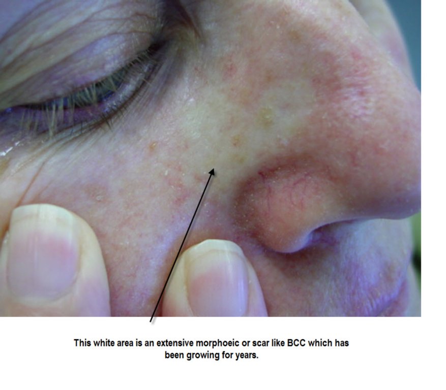
Electro desiccation can be used to treat superficial skin cancers. It is important to remember though those lesions that are not suitable for this treatment. This includes large basal cell or squamous cell skin cancers, including superficial types say over about 3cm in diameter, any morphoeic type BCC, any lesions that involve the nasal labial fold, the oral commisures, external auditory canal and inner canthus of the eye where the lesions can go deeper, any tumour that is recurrent or deeply invasive where there is surrounding scar formation as well would not be suitable for this technique. Also you should not use this technique on any type of suspected melanoma or pigmented lesion where excisional biopsy is the preferred method. When you are using electro desiccation on superficial basal or squamous cell skin cancers remember to treat at least a couple of millimetres beyond the apparent edge of the tumour and to make sure that you are getting any smaller areas that are not visible clinically. We generally talk of serial curettage. What this means is that you do at least two electro desiccations and curettes. Some people would suggest that you actually should do three but I usually find that this is unnecessary. Certainly two are required to adequately treat a lesion using this technique.


The one area that does give some complications using this technique is the scalp where occasionally granulation tissue will develop and it takes some time for it to settle down. Either a topical silver nitrate stick or some TCA can be applied on a weekly basis to reduce this granulation tissue until the lesion heals. Note that some patients though will develop a condition called erosive pustular dermatosis where granulation type tissue repeatedly occurs on the scalp and will in fact settle with the use of a topical steroid lotion such as Novasone lotion applied twice daily.
Electro desiccation over areas such as the anterior chest wall and the sternum or over the clavicles and deltoid regions the patient should be warned that keloid scarring may occur or at least hypertrophic scarring and this complication can be dealt with using intralesional steroid injections.


Spider angiomas can also be easily treated using electro desiccation but it requires only a very low setting to obliterate these lesions. They are best treated without local anaesthetic. You simply mark where the central vessel is and apply the needle tip at that point. The other vessels will collapse and settle if the central vessel is treated. You can though treat some of these larger tributary vessels radiating from the central vessels if necessary. For these the hyfrecator is gently moved along the line of the vessel with a linear motion. Sometimes Campbell de Morgan spots on the chest wall can be treated similarly but do require electro surgery in the desiccation mode to dry them out. They may need more than one treatment.
Electro cautery involves a different apparatus with a platinum tipped needle where the needle tip is heated by passing a current through it and this is then used to apply heat to an area and it is useful for reducing blood seepage over a larger area and for sealing any bleeding points after curettage. However a hyfrecator can also be used to take out the small individual bleeding points.
Variants of Simple Excisions
There are several variations of the fusiform ellipse and these include the curved ellipse, the lazy S excision and the M plasty. The M plasty is used where a fusiform ellipse would cross over the border of a cosmetic surgical unit. It is particularly useful if it is going to run toward the lip and cross the vermillion border or near an eyelid margin. Remember that a fusiform ellipse is usually drawn and done along the relaxed skin tension lines. There are various ways to start suturing an ellipse if you are using interrupted sutures. One method is the rule of halves, where the initial suture is placed in the centre of the lesion, and the defect on either side is gradually bisected with further sutures until full closure is achieved. Other people sometimes close the ends of an elipse first to get good closure here, and then subsequently close the rest of the ellipse.
Undermining
Undermining is an important technique used to free up the surrounding tissue and allow it to move easily into the defect. Most undermining should be done at the deep level of the excision to ensure that the base of the defect is adequately closed and there is no space left that could fill with blood or serum. Most undermining is best done with blunt tip scissors rather than with a blade. The scissors should be inserted in a closed fashion, and then opened to break down the tissue along the plane of separation.
A curved ellipse is sometimes necessary to close a defect on areas such as the cheeks. This occurs when you have different diameters of the two sides of the ellipse. It is best then closed by the rule of halves, allowing the two unequal sides to be closed. A lazy S closure of an ellipse creates two curves running in opposite directions. This helps reduce the puckering that can occur on convexities such as on extremities.
An M plasty is generally done to prevent an incision going into a significant cosmetic area and this is particularly an issue at the vermillion border or perhaps the eyebrows. Basically with an M plasty, you create two 30 degree angles at one end of the elipse and this shortens the length of the scar by about one quarter to one third. A three point stitch going through the bottom triangle tip is used to ensure adequate apposition of the tissue.
Healing by Secondary Intention
There are some situations where a wound may be left to heal by secondary intention. This can surprisingly be quite a successful technique to use. It is particularly used on the concave surfaces of the ears, and can also be used even on the medial epicanthus of the eye, provided there is an equal portion of the defect above and below the medial canthal opening. The concave surface of the ear is usually your best bet for this approach.
Listen to this lecture from MD Live on the basic elliptical excision and it's variations described above. Reference Only
Mohs Surgery, Slow Mohs, Frozen section control by Plastics
Mohs Surgery is used for recurrent and aggressive variants of BCC and SCC occuring in areas of the skin, particularly the face, where tissue sparing but adequate tumour clearance is of the essence. The tissue is incised not at 90 degrees but at 45 degrees sloping inwards. The reason for this is that the edges of the excised disc of tissue are then inked in 4 quadrants and turned down so that the complete outer edges of the disc and the base of the disc are in the same horizontal plane. This piece of tissue is then snap frozen and cut horizontally from the base up. Hence you have a piece of tissue with the base of the excised tumour in the centre surrounded on the outside by the four inked quadrants of the outer edges. Examining this allows you to see if there is any tumour centrally at the base of the excised specimen and the complete outer edges of the excised specimen. The different inked quadrant edges allow you to say where any edge recurrence is and it can then be re excised and processed similarly. By taking several cuts you can minimise the amount of tissue you need to excise to get complete tumour clearance both in depth and at all edges. The conventional vertical breadloafing of excised specimens misses much of the outer tumour edges.
Plastic surgeons who use frozen section control are still having their specimens breadloafed as hence the technique is inferior to Mohs.
Slow Mohs basically refers to excising a lesion one day and having it histologically examined without closing the defect until a day or so later when the specimen has been examined by H/E. Generally these specimens have breadloafing processing as well and hence fail to examine all the excised specimen edges.It is best to not use this term and instead refer to delayed closure after histological confirmation of margins.
Some other information on Moh's Surgery


When to consider referral of a patient elsewhere
1. Recurrent infiltrating BCCs
2. Aggressive poorly differentiated SCCs
3. Skin cancers involving the external auditory canal
4. Skin cancers involving the lips
5. Extensive scalp lesions
6. Skin cancers with perineural invasion and spread.
As a means of ending this general introduction to excisional surgery you should now listen to this lecture on cosmetic units on the face and how to avoid crossing over them in any excisional surgery you carry out. You will learn the different types of flap closures later on but look at this lecture from MD LIVE as a statement of general principles. Reference only at this stageMCQs for Module 1 Please attempt these before the second Wednesday teleconference for Module 1.
Also complete these Resource Assessment Questions for Module 1. These should be sent in before the second Wednesday Webinar on the Wednesday night. These are graded and count towards your assessment for the Course.
No comments:
Post a Comment